Arrived from Neighboring Country on Suspicion of Cancer, Patient Recovered and Left

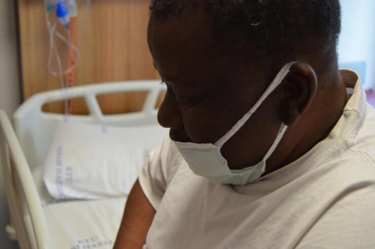
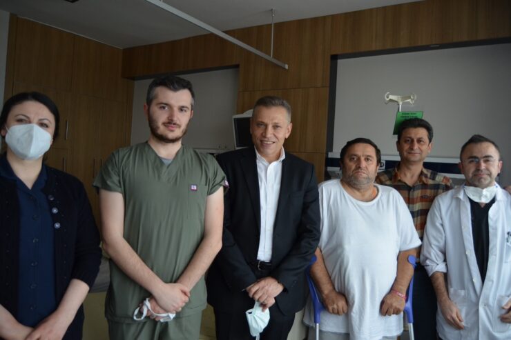
[vc_row][vc_column][vc_column_text]Nugzar Tsilosani, 50 years old, living in Georgia, went to a health center in his country after coughing up blood from his mouth. In the computerized tomography taken here, it was said that the lower part of the left lung did not swell and showed signs of destruction and it could be Lung Cancer. The patient came to Turkey for treatment and applied to Farabi Hospital of our University. In the examinations performed by Prof. Dr.  Celal TEKİNBAŞ and his team at Farabi Hospital. It was observed that the lower part of the left lung did not ventilate. Also an infection fluid empyema formed in the space between the ribs and the lung, and the meat structure, which we call parenchyma, was severely deteriorated. It was explained to the patient that this image could be seen not only in cancer but also in many other lung-related diseases, and the decision to operate was taken. 
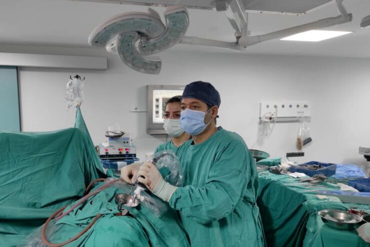
After the classical left posterolateral thoracotomy open surgery, the blood flow from the patient's mouth stopped with coughing and the shortness of breath improved. In the biopsy examination of the piece taken, it was understood that the patient did not have cancer, but that the structure of the lung trachea, called bronchiectasis, was severely deteriorated, and that it was a disease caused by the decay of the meat part of the lung where gas exchange takes place. 
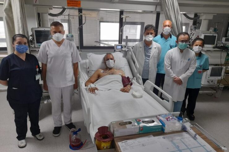
The patient, who was discharged, returned to his country with the happiness of both regaining his health and learning that he did not have cancer.[/vc_column_text][/vc_column][/vc_row][vc_row][vc_column][vc_gallery type="image_grid" images="5172,5173,5174,5175,5176"][/vc_column][/vc_row]

Patient Started Walking Again With the Operation Performed in Farabi

[vc_row][vc_column][vc_column_text]A 52-year-old male patient named Fousseini DIAKITE was admitted to KTU Farabi Hospital from Mali, a West African country, with complaints of inability to walk and severe pain in the left leg. The patient stated that many traditional methods had been tried in his country before, but his complaints were getting worse and the scars seen in the lumbar region during his examination belonged to previous injections he had had. When the magnetic resonance imaging of the patient for spinal cord diseases was evaluated, there were observed egenerative severe stenosis in the cervical spinal canal and infection involving the spine and intervertebral joints, which were thought to have occurred due to injections in the lumbar region,. Patient was taken into neck and lumbar spine operations simultaneously by Prof. Dr. Ertuğrul ÇAKIR and his team, in Brain and Nerve Surgery Clinic,. After the operation, it was observed that the pain in the left leg of the patient decreased and he was able to walk without support in the early period. Antibiotherapy was started for the infection in the lumbar spine. Although the patient's infection is under control, his treatment is still ongoing. Prof. Dr. Ertuğrul ÇAKIR gave the following information about the operation: “When our patient applied to our clinic, he had severe pain and I learned that he could not walk for 1 year. I examined the old-style MR (EMAR) films in their file, but there was no explanatory image. I suggested medicated MRI (EMAR) of the spinal cord, as I thought it would explain the current clinic. When the patient came with the results of the examination, I saw that there were severe stenosis at different levels due to degeneration in the cervical spinal canal, and that there were signs in favor of the infection between the lumbar spine, which I thought was caused by the scar in the lumbar region that I had noticed before. Since these two diseases require surgery, my team and I performed decompression and posterior instrumentation surgeries on the neck and waist regions of the patient.” Prof. Dr. Ertuğrul ÇAKIR said the following about the operation and diseases: “There are three main reasons for the narrowing of the cervical spinal canal. These can be listed as structural stenosis, stenosis due to aging and wear in the disc, and stenosis due to aging and wear in the spine joints. In general, it is seen as cervical myelopathy findings (limb dysfunction in arms and legs, gait disturbances, bladder and bowel dysfunction, etc.) and cervical radiculopathy (pain, numbness, loss of function in arms and legs, etc.). Although partial benefit can be obtained with drug treatment, surgery has an important place in advanced and severe cases.’’ “Lumbar spondylodiscitis and spine infections generally present with severe pain and loss of limb function, and surgery is performed when drug therapy alone is not sufficient. Although satisfactory results are not always obtained after the surgery, depending on the severity of the spinal cord and nerve damage, we are very happy that many of our patients benefited from the surgery and started walking again, as in this patient example.’’[/vc_column_text][/vc_column][/vc_row][vc_row][vc_column][vc_gallery type="image_grid" images="5146,5145,5147,5148,5149,5150,5151,5152,5153,5154,5155,5156"][/vc_column][/vc_row]

Healing Hand Extending from Trabzon to Africa, The Mass in the Brain of the Patient Coming from Mali was Successfully Removed

[vc_row][vc_column][vc_column_text]A 60-year-old male patient named Daff Tidiani applied to KTU Farabi Hospital from Mali, a West African country, with complaints of blurred vision and headache. It was learned that the patient had previously undergone nasal pituitary gland surgery in a clinic in Tunisia. The patient stated that he applied to KTU Farabi Hospital Brain and Nerve Surgery Clinic because of the complaint of blurred vision despite the surgery he had done. In the neurological and eye examinations performed in the clinic, the patient had visual loss and a recurrent mass (macroadenoma) pressing on the optic nerves was detected in the pituitary MRI (Emar). After the patient was hospitalized and the necessary examinations were made, he was taken into surgery. Surgery it was made by the team consisting of; Prof. Dr. Erhan ARSLAN from the Department of Neurosurgery. and Prof. Dr. Osman BAHADIR from the Department of Otorhinolaryngology. In the surgery, the tumor in the pituitary gland was successfully removed under the microscope by entering through the nose (endoscopic endonasal transsphenoidal approach). The patient, whose vision problems improved after surgery, was discharged with full recovery.   Prof. Dr. Erhan ARSLAN gave the following information about this disease and surgery: Pituitary adenomas are mostly benign tumors and arise from the pituitary gland located in the sella turcica at the base of the skull. The pituitary gland is a 1 cm-sized organ located alone behind the eyes and responsible for hormone production. The pituitary gland is a central and vital organ that ensures the secretion of many hormones in our body and the water balance of the body. The clinical manifestations of pituitary adenomas basically vary depending on the pituitary cell from which they originate, whether they produce hormones or not, and the size of the tumor they reach. Accordingly, adenomas  smaller than 1 cm in diameter are called microadenomas; Adenomas larger than 1 cm are called macroadenomas. In pituitary gland surgeries, the most important structure to be considered while removing the tumor (macroadenoma) is the Internal Carotid arteries. Injury to the Internal Carotid arteries, known as the carotid artery from the neck, can have serious consequences, including paralysis and death.[/vc_column_text][/vc_column][/vc_row][vc_row][vc_column][vc_gallery type="image_grid" images="4755,4756,4757,4758,4759"][/vc_column][/vc_row]

Came from Baghdad, Regained His Health in Trabzon

[vc_row][vc_column][vc_column_text]After our hospital received the Health Tourism Authorization Certificate, an important operation was performed on the first international patient. The surgery, classified as major (group A), was performed on a 45-year-old patient named Mahmood FAKHRI, who came from Iraq. After the examinations performed on the 45-year-old father of 2 children, who came from Baghdad, the capital of Iraq, who was diagnosed with an esophageal tumor, the esophageal tumor surgery, which lasted about 5 hours, was successfully performed. The treatment of the patient, who was taken to the intensive care unit after the operation, is still continuing. The surgery was performed by the team including Prof. Dr. Atila TÜRKYILMAZ and Dr. Alaattin BURAN  under the management of KTU Farabi Hospital Chief Physician Prof. Dr. Celal TEKİNBAŞ. Prof. Dr. Celal TEKİNBAŞ said, “After receiving the health tourism authorization certificate, we started to take steps that could lead to very important gains for our city, region and country. In this context, we performed a successful operation that lasted about 5 hours for our patient who came to our hospital from Iraq the previous day due to an esophageal tumor. There was no problem in the post-operative follow-up of our patient and we continue to follow up. We plan to discharge our patient after 4-5 days. I wish this step we have taken to be a start for all patients from near and far countries who need healing.” KTU Farabi Hospital teams, who have performed many difficult surgeries like this until now, are determined to transfer this great experience and competence they have to international patients coming within the scope of Health Tourism as a cure.[/vc_column_text][/vc_column][/vc_row][vc_row][vc_column][vc_gallery type="image_grid" images="4554,4555,4556,4557,4558,4559"][/vc_column][/vc_row]

Our Hospital Received Health Tourism Authorization Certificate

[vc_row][vc_column][vc_column_text]Our hospital has been awarded a health tourism authorization certificate. Thanks to this document, there are no obstacles in front of us regarding health tourism activities. In this way, we think that the start of international patient admission within the scope of health tourism of our hospital will make important financial contributions to both our city, region and country and will become an international healing center. We believe that we will get significant distances in a short time thanks to the determination of our Rector Prof. Dr. Hamdullah ÇUVALCI and our Chief Physician Prof. Dr. Celal TEKİNBAŞ to improve the necessary infrastructure in this regard every day.[/vc_column_text][/vc_column][/vc_row][vc_row][vc_column][vc_gallery type="image_grid" images="4549,4550,4551"][/vc_column][/vc_row]

Bpth COVID-19 And CCHF Diseases Caught

[vc_row][vc_column][vc_column_text]The 60-year-old ID came to the Medical Faculty Hospital of the Karadeniz Technical University (KTU) Farabi with complaints of fever, weakness and shortness of breath. The patient named Covid-19 and Crimean Congo Hemorrhagic Fever (CCHF) was diagnosed, treated and discharged. Experts stated that the coexistence of Covid-19 and CCHF diseases is a very rare situation in our country, while such cases will gradually take their place in the literature. Comment on the subject Technical University Karadeniz (KTU) Medical Faculty Head of the Department of Infectious Diseases and Clinical Microbiology Prof. DR. Selcuk Kaya, said that with the emergence of tick cases, they are now carefully examining the patients who come to their service thinking they could have both CCHF and Covid-1 9 cases as they show similar symptoms. “THE KKKA IN THE REGION COMES TO US BY SLOWLY INCREASING ITS FREQUENCY” As a result of these careful examinations, the 60-year-old ID came to them 10 days ago with complaints of fever, weakness and shortness of breath. Noting that they have diagnosed both Covid-19 and Crimean Congo Hemorrhagic Fever (CCHF), Prof. DR. Selçuk Kaya said, “We are experiencing a very serious period of pandemic worldwide. We see very serious consequences in our country of this virus which has caused the death of many people. But of course infectious diseases, which we call many other endemics that we are used to seeing frequently in our country and region, also retain their significance. As a result, the diseases did not stop. Especially at the beginning of the summer months, people went to open areas and met us, which gradually increased the incidence of CCHF in the area due to the increased contact with nature. In this context, we have admitted a patient aged 60 with a preliminary diagnosis of Covid-19 to the Covid service. The patient came to us with a fever, weakness and some breathing problems. As we are an endemic region, we always think of the cases of Crimean-Congo Hemorrhagic Fever, especially those that come from the high parts of Gümüşhane, Bayburt and Giresun. Based on the positive CCHF tests in the tests we carried out for this purpose, we diagnosed the case as both a Covid and CCHF infection case and carried out treatments. First we planned treatments related to Covid, then we put the appropriate supportive treatments on the agenda after the positive CCHF test. After a treatment period of approx. 10 days, the patient gradually recovered from his blood count and general clinical picture and, luckily, was successfully treated and discharged in our service without intensive medical care and support such as dialysis. “I CANNOT SAY IT WAS A SURPRISE, IT IS IMPORTANT THAT IT WAS DIAGNOSTED” Kaya said such cases are rare, Kaya said: “It is no surprise to see Covid-19 patients during the pandemic and due to increasing tick contact with the warming weather, our region is one of the places where tick cases are occurring are most common. Hence, we need to keep an eye on the cases that come from risky locations in the eastern Black Sea region. Actually, I can’t say it was a surprise. It is important that it be diagnosed. Because if we hadn’t labeled these cases as Covid cases during the pandemic we were going through and carefully examined them for other CCHF and other diseases, it could have been overlooked. In Turkey, such cases are both theoretically and practically possible. I haven’t heard such a thing outright, but; I think that in these and similar cases we also found something positive in our 50-year-old patient. Here, too, the focus is on fever and malaise. The general clinical picture is encouraging. We’ll look into their tests in more detail. The arrival of these and similar cases may gradually take their place in the literature. At the moment we can say that these are rare cases, “he said.[/vc_column_text][/vc_column][/vc_row][vc_row][vc_column][vc_gallery type="image_grid" images="4543,4546,4541,4542,4544,4545"][/vc_column][/vc_row]

Tracheal Stenosis Caused By Mechanical Ventilation Treatment Was Removed Surgically

[vc_row][vc_column][vc_column_text]Mechanical ventilator-associated tracheal stenosis was successfully treated with surgery in the patient who was treated in the intensive care unit due to covid 19 infection. A 52-year-old female patient, who was hospitalized in the intensive care unit due to the COVID-19 infection and was discharged after being treated with a mechanical ventilator, applied to the Thoracic Surgery clinic after 1 month with complaints of shortness of breath. As a result of detailed examinations, a 4.5 cm long stenosis was detected in the trachea of ​​our patient. The stenosis in the trachea was enlarged by the Rigid Bronchoscopy procedure to open the airway. The respiratory distress of the patient was temporarily relieved and a detailed diagnosis of the problematic area was provided. As a result of the evaluations, operation was planned for the narrowed trachea 2 weeks later. In the surgery performed by the team led by our Chief Physician Prof. Dr.  Celal TEKİNBAŞ, the narrowed part of the trachea of ​​the patient was cut and removed, and the healthy ends were stitched together. Our patient, who was hospitalized for only 5 days after the operation, was discharged with full recovery. There is no problem in our patient in the next 10-day and 2-month controls.[/vc_column_text][vc_single_image image="4536" img_size="large" alignment="center"][vc_column_text]During the pandemic period, the number of patients who received respiratory support with a mechanical ventilator increased. Trachea stenosis may develop in approximately 5% of patients who receive this treatment. In these cases, the definitive treatment is with the surgery performed by stitching the two healthy ends together after cutting and removing the stenosis part of the trachea. KTÜ Farabi Hospital is an experienced center that successfully treats these cases.[/vc_column_text][/vc_column][/vc_row][vc_row][vc_column][vc_gallery type="image_grid" images="4538,4537,4536,4535"][/vc_column][/vc_row]

The Deadly Cyst in the Heart Has Been Successfully Removed!

[vc_row][vc_column][vc_column_text]A 34-year-old patient named B. B had a cyst in his heart in the echocardiography taken at Artvin State Hospital, where he went to get a medical report to get a job in December. The patient, who was urgently referred to our hospital for advanced treatment, was treated in the Department of Cardiovascular Surgery of our hospital. In the examinations performed, it was determined that the cyst located in  the heart developed due to the parasite named "echinococcus" which is transmitted from animals to humans. It was decided to remove the cyst, which was observed to be quite enlarged in the heart and almost completely filled the heart cavity, with open heart surgery because it was life-threatening. The patient, whose heart cyst was removed after a successful operation, was discharged home 4 days after the operation. During his controls, it was seen that the problem in his heart was completely cured. Prof. Dr. Ahmet Coşkun ÖZDEMİR stated that cyst transmitted from animals to humans through some parasites is not uncommon. However, he stated that these cysts are generally located in the liver and lungs and that these cysts are seen in the heart in only 1% of the patients. Prof. Dr. Ahmet Coşkun ÖZDEMİR stated that the cyst was located in the right ventricle of the heart in this patient and therefore this case is a very rare condition in the world. Stating that the patient's problems were completely resolved after the surgery and he was discharged after 4 days, Prof. Dr. ÖZDEMİR  said that“The patient was living with a fatal disease in his heart, although he was not aware of it at all. Until today, it has been a great chance for the patient that the cyst has not ruptured. He gained his health with surgery, ” adding that people dealing with animal husbandry should be careful about hydatid cyst disease.[/vc_column_text][vc_single_image image="4496" img_size="large" alignment="center"][vc_column_text]Assistant Professor Dr. İlke Onur KAZAZ, stated that the treatment of metastatic kidney tumors with open and closed surgery methods was performed in Farabi Hospital, Department of Urology and that a positive contribution was made to the life expectancy of patients. Dr. Kazaz added that KTU Farabi Hospital, Clinic of Urology has successfully performed many similar operations thanks to its advanced technology and experienced surgeons.[/vc_column_text][/vc_column][/vc_row][vc_row][vc_column][vc_gallery type="image_grid" images="4503,4504,4505,4506,4507,4502"][/vc_column][/vc_row]

The Patient With Giant Kidney Mass Regained His Health

[vc_row][vc_column][vc_column_text]Another critical operation was successfully carried out at KTU Farabi Hospital. 58-year-old male patient named S.A admitted to Urology clinic. In the examinations, a 35-centimeter mass was detected in the right kidney. It was observed that the mass extended to other internal organs in the abdominal cavity. The right kidney of the patient, who was decided to be operated, was removed together with the mass. Assistant Professor Dr. İlke Onur KAZAZ, Assistant Professor Dr. Fatih ÇOLAK and their team who carried out the operation stated that the operation took about 3 hours and was successfully concluded. The patient was discharged with full recovery after a one-week follow-up.[/vc_column_text][vc_single_image image="4496" img_size="large" alignment="center"][vc_column_text]Assistant Professor Dr. İlke Onur KAZAZ, stated that the treatment of metastatic kidney tumors with open and closed surgery methods was performed in Farabi Hospital, Department of Urology and that a positive contribution was made to the life expectancy of patients. Dr. Kazaz added that KTU Farabi Hospital, Clinic of Urology has successfully performed many similar operations thanks to its advanced technology and experienced surgeons.[/vc_column_text][/vc_column][/vc_row][vc_row][vc_column][vc_gallery type="image_grid" images="4499,4498,4497,4495,4496"][/vc_column][/vc_row]